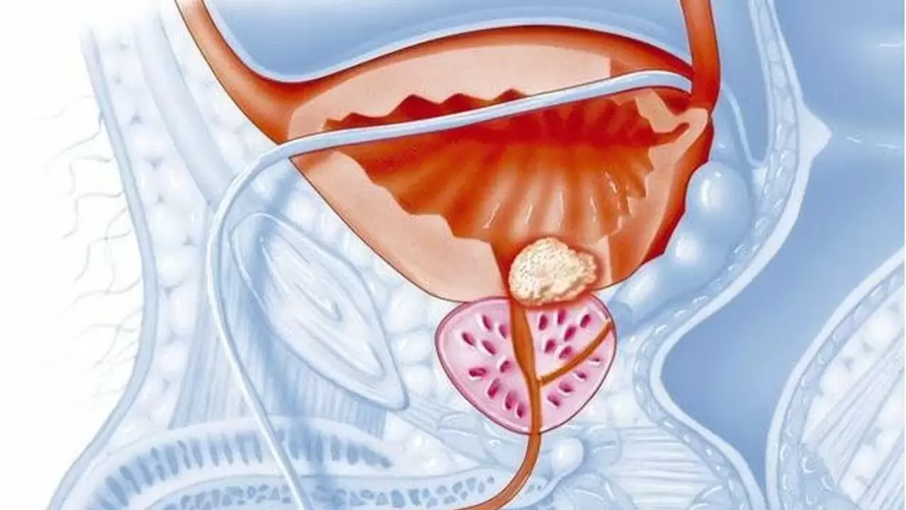
Chronic prostatitis – inflammatory disease of the prostate gland of various etiologies (including non-infectious), manifested by pain or discomfort in the pelvic area and urinary disturbances for 3 months or more.

I. Introduction section
Protocol name: Inflammatory disease of the prostate gland
Protocol code:
ICD-10 codes:
N41. 0 Acute prostatitis
N41. 1 Chronic prostatitis
N41. 2 Prostatic abscess
N41. 3 Prostatocystitis
N41. 8 Other inflammatory diseases of the prostate gland
N41. 9 Inflammatory disease of the prostate, unspecified
N42. 0 Prostate stones
Prostate stones
N42. 1 Congestion and bleeding in the prostate gland
N42. 2 Prostate atrophy
N42. 8 Other specific diseases of the prostate gland
N42. 9 Diseases of the prostate gland, unspecified
Abbreviations used in the protocol:
ALT - alanine aminotransferase
AST - aspartate aminotransferase
HIV - human immunodeficiency virus
ELISA - enzyme immunoassay
CT - computed tomography
MRI - magnetic resonance imaging
MSCT - multi-slice tomography
DRE - digital rectal examination
PSA - prostate specific antigen
DRE - digital rectal examination
PC - prostate cancer
CPPS - chronic pelvic pain syndrome
TUR - transurethral resection of the prostate gland
Ultrasound - ultrasound examination
ED - erectile dysfunction
ECG - electrocardiography
IPSS - International Prostate Symptom Score (international symptom index for prostate disease)
NYHA – New York Heart Association
Protocol development date: 2014
Patient category: men of reproductive age.
User protocol: andrologists, urologists, surgeons, therapists, general practitioners.
Level of Evidence
Level |
Types of evidence |
| 1a | Evidence comes from meta-analyses of randomized trials |
| 1b | Evidence from at least one randomized trial |
| 2a | Evidence comes from at least one well-designed, controlled, non-randomized trial |
| 2b | Evidence comes from at least one well-designed, controlled, quasi-experimental study |
| 3 | Evidence obtained from well-designed non-experimental research (comparative research, correlational research, analysis of scientific reports) |
| 4 | Evidence is based on expert opinion or experience |
Degree of recommendation
| A | Results are based on homogeneous, high-quality, problem-specific clinical trials, with at least one randomized trial |
| INSIDE | Results obtained from well-designed, non-randomized clinical studies |
| WITH | No clinical studies of sufficient quality have been conducted |
Classification
Clinical classification
Classification of prostatitis (National Institute of Health (NYHA), United States, 1995)
Category I - acute bacterial prostatitis;
Category II – chronic bacterial prostatitis, found in 5-10% of cases; Category III - chronic abacterial prostatitis/chronic pelvic pain syndrome, diagnosed in 90% of cases;
Subcategory III A – chronic inflammatory pelvic pain syndrome with increased leukocytes in prostatic secretions (more than 60% of all cases); Subcategory III B – CPPS - chronic non-inflammatory pelvic pain syndrome (without an increase in leukocytes in prostate secretion (about 30%));
Category IV – asymptomatic inflammation of the prostate, detected during examination for other diseases, based on the results of the analysis of prostate secretion or its biopsy (histological prostatitis unknown);
Diagnostics
II. Methods, approaches and procedures for diagnosis and treatment
List of basic and additional diagnostic steps
Basic (mandatory) diagnostic examinations are performed on outpatients:
- collection of complaints, medical history;
- digital rectal examination;
- fill out the IPSS questionnaire;
- ultrasound examination of the prostate;
- prostatic secretions;
Additional diagnostic tests performed on outpatients: prostatic secretions;
The minimum list of examinations that must be carried out when referring for planned hospitalization:
- general blood test;
- general urinalysis;
- biochemical blood tests (determination of blood glucose, bilirubin and fractions, AST, ALT, thymol test, creatinine, urea, alkaline phosphatase, blood amylase);
- microreactions;
- coagulogram;
- HIV;
- ELISA for viral hepatitis;
- fluorography;
- ECG;
- blood group.
Basic (mandatory) diagnostic examinations are carried out at the hospital level:
- PSA (total, free);
- bacteriological culture of prostate secretion obtained after massage;
- transrectal ultrasound examination of the prostate;
- bacteriological culture of prostate secretion obtained after massage.
Additional diagnostic tests carried out at the hospital level:
- uroflowmetry;
- cystotonometry;
- MSCT or MRI;
- urethrocystoscopy.
(level of evidence - I, strength of recommendation - A)
Diagnostic measures carried out at the emergency level: not carried out.
Diagnostic criteria
Complaints and anamnesis:
Complaint:
- pain or discomfort in the pelvic area that lasts for 3 months or more;
- Frequent localization of pain is the perineum;
- discomfort may be suprapubic;
- discomfort in the groin and pelvis;
- discomfort in the scrotum;
- discomfort in the rectum;
- discomfort in the lumbosacral region;
- pain during and after ejaculation.
Anamnesis:
- sexual dysfunction;
- libido suppression;
- deterioration in the quality of spontaneous and/or sufficient erection;
- premature ejaculation;
- in the final stages of the disease, slow ejaculation;
- "erasing" the emotional coloring of orgasm.
The impact of chronic prostatitis on quality of life, according to the unified quality of life rating scale, is comparable to the impact of myocardial infarction, angina pectoris and Crohn's disease. (level of evidence - II, strength of recommendation - B).
Physical examination:
- swelling and tenderness of the prostate gland;
- enlargement and smoothing of the median groove of the prostate gland.
Laboratory research
To increase the reliability of laboratory test results, they should be carried out before the appointment or 2 weeks after the end of taking antibacterial agents.
Microscopic examination of prostate secretions:
- determination of the number of leukocytes;
- determination of the amount of lecithin grains;
- determination of the number of amyloid bodies;
- determination of the number of Trousseau-Lallemand bodies;
- determination of the number of macrophages.
Bacteriological study of prostate secretion: determining the nature of the disease (bacterial or abacterial prostatitis).
Criteria for bacterial prostatitis:
- the third part of urine or prostatic secretion contains bacteria of the same strain in a titer of 103 CFU/ml or more, provided that the second part of urine is sterile;
- a tenfold or greater increase in bacterial titers in the third portion of urine or in prostatic secretions compared to the second portion;
- the third part of urine or prostate secretion contains more than 103 CFU/ml of true uropathogenic bacteria, different from other bacteria in the second part of urine.
The main importance in the occurrence of chronic bacterial prostatitis of gram-negative microorganisms from the Enterobacteriaceae family (E. coli, Klebsiella spp, Proteus spp, Enterobacter spp, etc. ) and Pseudomonas spp, as well as Enerococcus faecalis has been proven.
Blood sampling to determine serum PSA concentration should be performed no earlier than 10 days after DRE. Prostatitis can cause an increase in PSA concentration. However, when the PSA concentration exceeds 4 ng/ml, the use of additional diagnostic methods, including prostate biopsy, is indicated to exclude prostate cancer.
Instrumental studies:
Transrectal ultrasound of the prostate gland: for differential diagnosis, to determine the form and stage of the disease with subsequent monitoring throughout treatment.
Ultrasound: assessment of the size and volume of the prostate, echostructure (cysts, stones, fibrous-sclerotic changes in the organ, prostate abscess). Hypoechoic areas in the peripheral zone of the prostate are suspicious for prostate cancer.
X-ray study: with diagnosed bladder outlet obstruction to clarify the cause and determine further treatment tactics.
Endoscopic methods (urethroscopy, cystoscopy): carried out according to strict indications for the purpose of differential diagnosis, including broad-spectrum antibiotics.
Urodynamic study (uroflowmetry): determination of urethral pressure profile, pressure/flow study,
Cystometry and pelvic floor muscle myography: if bladder outlet obstruction is suspected, which often accompanies chronic prostatitis, as well as neurogenic disorders of urination and pelvic floor muscle function.
MSCT and MRI of pelvic organs: for differential diagnosis with prostate cancer.
Tips for consulting a specialist: consultation with an oncologist - if PSA is more than 4 ng/ml, to exclude malignant prostate formation.
Differential diagnosis
Differential diagnosis of chronic prostatitis
For the purpose of differential diagnosis, the condition of the rectum and surrounding tissues should be evaluated (level of evidence - I, strength of recommendation - A).
Nosologies |
Characteristic syndromes/symptoms | Differential test |
| Chronic prostatitis | The average age of the patients was 43 years. Pain or discomfort in the pelvic area that lasts for 3 months or more. The most common localization of pain is the perineum, but discomfort can be in the suprapubic, pelvic inguinal, as well as in the scrotal, rectal, and lumbosacral regions. Pain during and after ejaculation. Urinary dysfunction often manifests itself as an irritating symptom, less often as a symptom of bladder outlet obstruction. |
CURRENT - you can detect swelling and tenderness of the prostate gland, and sometimes enlargement and smoothness of the median groove. For the purpose of differential diagnosis, the condition of the rectum and surrounding tissues should be evaluated. Prostatic secretions - determine the number of leukocytes, lecithin granules, amyloid bodies, Trousseau-Lallemand bodies and macrophages. Bacteriological study of prostate secretions or urine obtained after the massage is carried out. Based on the results of this study, the nature of the disease is determined (bacterial or abacterial prostatitis). Criteria for bacterial prostatitis
Ultrasound of the prostate gland in chronic prostatitis has high sensitivity but low specificity. This study allows not only to carry out differential diagnosis, but also to determine the form and stage of the disease with subsequent monitoring throughout the treatment. Ultrasound makes it possible to assess the size and volume of the prostate, echostructure |
| Benign prostatic hyperplasia (prostatic adenoma) | It is observed more often in people over the age of 50. A gradual increase in urination and a slow increase in urinary retention. Increased frequency of urination is typical at night (for chronic prostatitis, increased frequency of urination during the day or in the early morning). | PRI - the prostate gland is painless, enlarged, densely elastic, the central groove is smoothed, the surface is smooth. Prostatic secretion - the amount of secretion increased, but the number of leukocytes and lecithin grains remained within the physiological norm. The secretory reaction is neutral or slightly alkaline. Ultrasound - deformation of the bladder neck is observed. Adenoma protrudes into the bladder cavity in the form of a bright red, lumpy formation. There is significant proliferation of glandular cells in the cranial part of the prostate gland. The structure of the adenoma is homogeneous with dark areas of normal shape. There is an increase in the gland in the anteroposterior direction. With fibroadenoma, a bright echo from the connective tissue is detected. |
| Prostate cancer | People over the age of 45 are affected. When diagnosing chronic prostatitis and prostate cancer, there is a similar localization of pain. Pain in prostate cancer in the lumbar region, sacrum, perineum, and lower abdomen can be caused by both processes in the gland itself and by metastases in the bones. Rapid development of complete urinary retention often occurs. Severe bone pain and weight loss may occur. | IF - nodes of individual cartilaginous density or lumpy dense infiltration of the entire prostate gland are determined, which are limited or spread to the surrounding tissue. The prostate gland does not move, does not hurt. PSA - more than 4. 0 ng/ml Prostate biopsy - a collection of malignant cells in the form of a ductal cast is determined. Atypical cells are characterized by hyperchromatism, polymorphism, variability in size and shape of nuclei, and mitotic figures. Cystoscopy - a pale pink mass is determined, surrounding the bladder neck in a ring (the result of infiltration of the bladder wall). Often swelling, hyperemia of the mucous membrane, malignant proliferation of epithelial cells. Ultrasound - asymmetry and enlargement of the prostate gland, significant deformation. |
Treatment
Treatment goals:
- elimination of inflammation in the prostate gland;
- relieve symptoms of exacerbation (pain, discomfort, urination and sexual dysfunction);
- prevention and treatment of complications.
Treatment tactics
Non-drug treatment:
Diet No. 15.
Mode: general.
Drug treatment
When treating chronic prostatitis, it is necessary to simultaneously use several drugs and methods that act on different parts of the pathogenesis and allow the elimination of infectious agents, normalization of blood circulation in the prostate, adequate drainage of prostatic acini, especially in the peripheral zone, normalization of important hormone levels andimmune response. Antibacterial drugs, anticholinergics, immunomodulators, NSAIDs, angioprotectors, vasodilators, prostate massage are recommended, and therapy with alpha blockers is also possible.
Other treatments
Other types of treatment provided on an outpatient basis:
- transrectal microwave hyperthermia;
- physiotherapy (laser therapy, mud therapy, phono-electrophoresis).
Other types of services provided at the stationary level:
- transrectal microwave hyperthermia;
- physiotherapy (laser therapy, mud therapy, phono-electrophoresis).
Other types of treatment provided at the emergency level: not provided.
Surgical intervention
Surgical intervention provided on an outpatient basis: not performed.
Surgical intervention is provided in an inpatient setting
Type:
Transurethral incision at 5, 7, and 12 o'clock.
Hint:
carried out in a hospital setting if the patient has prostate fibrosis with a clinical picture of bladder outlet obstruction.
Type:
Transurethral resection
Hint:
used for calculous prostatitis (especially when stones are localized that cannot be treated conservatively in the central, temporal and periurethral zones).
Type:
Resection of the spermatic tubercle.
Hint:
with sclerosis of the seminal tubercles, accompanied by obstruction of the ejaculatory ducts and prostatic excretion.
Preventive measures:
- abandon bad habits;
- eliminate the influence of harmful influences (cold, physical inactivity, prolonged sexual abstinence, etc. );
- diet;
- spa treatments;
- normalization of sexual life.
Further management:
- observation by a urologist 4 times a year;
- Ultrasound of the prostate and residual urine in the bladder, DRE, IPSS, prostate secretion 4 times a year
Indicators of treatment effectiveness and safety of the diagnostic and treatment methods described in the protocol:
- absence or reduction of characteristic complaints (pain or discomfort in the pelvis, perineum, suprapubic area, pelvic inguinal area, scrotum, rectum);
- reduction or absence of swelling and tenderness of the prostate gland according to DRE results;
- reduction of inflammatory indicators of prostate secretion;
- reduction of swelling and prostate size according to ultrasound.


